


01 Oct Electron microscope analyses
Two basic types of methods are possible to apply in a frame of the electron microscopy. Each of these methods use different equipment and is based on different attitude and sample pre-treatment for the analysis and following outputs.
Scanning electron microscopy (SEM) using the JEOL 6490 LV electron microscope is a non-destructive or semi-destructive method suitable especially on the samples, where any kind of a damage or an adjustment before the analysis is acceptable. SEM enables to analyse either natural inorganic materials or artificially made materials (minerals, rocks, metals, glasses, slags, concretes etc.) on samples up to 6 cm high and up to 10 cm wide respectively. Lower-pressure mode (suitable e.g. for porous materials) as well as no necessity of an application of a conductive layer on the sample surface (atomic carbon or gold) are important is an advantage of SEM. On the other hand, if necessary, it is also possible to apply the conductive layer to study a micro-relief of the sample surface. It is also feasible to determine the elemental composition of analysed material with the use of the EDX analysis (Energy-dispersive-X-ray-spectroscopy), which enables to detect presence of major chemical elements in the material studied with an accuracy up to 0.X wt.% (elements with the atomic mass higher than Fluor). Chemical analyses can be performed from a surface area of a few square micrometres to circa one square millimetre.
The electron microprobe CAMECA SX 100 is a second equipment using electron microscopy. However, according to the application possibilities, it is necessary to prepare a thin polished section (0.03-0.1 mm thick sample circa 2,5 x 3 cm glued to glass plate) or a polished section in an epoxide tablet with a diameter of 2 cm (suitable also for powders or other incoherent materials). The analyses are performed in the mode of the high vacuum, which brings, apart from a necessity of application of conductive layer (atomic carbon), also chemical measurement accuracy improvement (up to hundreds of ppm). In the chemical elemental analysis, it is possible to achieve the accuracy about hundreds of ppm. The WDX method (Wave-length dispersive X-ray spectroscopy) is applied however, it is also possible to use the EDX analysis, which can help with a swift identification of major, minor and trace minerals and phases in rocks and other inorganic materials. Apart of the spot analyses (usually 1-10 µm) it is also possible to apply sectional analyses or elemental distribution maps.
Both methods allow graphical recording in the form of BSE images (Back-scattered electrons), which perfectly show phase differences among individual grains of different chemical composition and also a zoning or other heterogeneities or SE (secondary electrons), enable to display a detailed morphology of a micro-relief of a sample surface.
For both methods (SEM and the electron microprobe), we offer the possibility of sample preparation depending on the use and need.
In the ERCA Centre the electron microscope analyses are guaranteed by a specialist Mgr. Petr Gadas, PhD., and they will be performed in the laboratory of electron microscopy and microanalyses of the Faculty of Science of the Masaryk university in Brno.
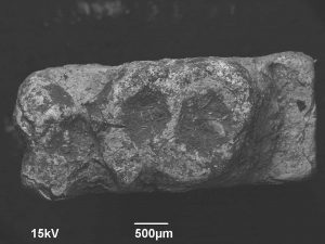
Photograph of the Early Modern Age letter in a combination of reflected and secondary electrons, which enables an anylysis of the alloy composition as well as of a character of a surface relief.
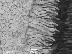
BSE photograph of a surface of Medieval glaze on a shard surface, which documents a manner and an extent of its degradation.